A lire sur: http://www.technologyreview.com/featuredstory/526501/brain-mapping/
A new map, a decade in the works, shows structures of the brain in far greater detail than ever before, providing neuroscientists with a guide to its immense complexity.
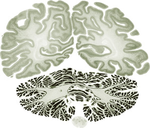
A section of the human brain map created by a team of international researchers shows details as small as 20 micrometers.
A new map, a decade in the works, shows structures of the brain in far greater detail than ever before, providing neuroscientists with a guide to its immense complexity.
Breakthrough
A high-resolution map that shows structures of the human brain as small as 20 micrometers.
Why It Matters
As neuroscientists try to understand how the brain works, they need a detailed map of its anatomy.
Key Players
- Katrin Amunts, Jülich Research Centre
- Alan Evans, Montreal Neurological Institute
- Karl Deisseroth, Stanford University
A section of the human brain map created by a team of international researchers shows details as small as 20 micrometers.
Neuroscientists have made remarkable progress in recent years toward understanding how the brain works. And in coming years, Europe’s Human Brain Project will attempt to create a computational simulation of the human brain, while the U.S. BRAIN Initiative will try to create a wide-ranging picture of brain activity. These ambitious projects will greatly benefit from a new resource: detailed and comprehensive maps of the brain’s structure and its different regions.
As part of the Human Brain Project, an international team of researchers led by German and Canadian scientists has produced a three-dimensional atlas of the brain that has 50 times the resolution of previous such maps. The atlas, which took a decade to complete, required slicing a brain into thousands of thin sections and digitally stitching them back together with the help of supercomputers. Able to show details as small as 20 micrometers, roughly the size of many human cells, it is a major step forward in understanding the brain’s three-dimensional anatomy.
To guide the brain’s digital reconstruction, researchers led by Katrin Amunts at the Jülich Research Centre in Germany initially used an MRI machine to image the postmortem brain of a 65-year-old woman. The brain was then cut into ultrathin slices. The scientists stained the sections and then imaged them one by one on a flatbed scanner. Alan Evans and his coworkers at the Montreal Neurological Institute organized the 7,404 resulting images into a data set about a terabyte in size. Slicing had bent, ripped, and torn the tissue, so Evans had to correct these defects in the images. He also aligned each one to its original position in the brain. The result is mesmerizing: a brain model that you can swim through, zooming in or out to see the arrangement of cells and tissues.
At the start of the 20th century, a German neuroanatomist named Korbinian Brodmann parceled the human cortex into nearly 50 different areas by looking at the structure and organization of sections of brain under a microscope. “That has been pretty much the reference framework that we’ve used for 100 years,” Evans says. Now he and his coworkers are redoing Brodmann’s work as they map the borders between brain regions. The result may show something more like 100 to 200 distinct areas, providing scientists with a far more accurate road map for studying the brain’s different functions.
“We would like to have in the future a reference brain that shows true cellular resolution,” says Amunts—about one or two micrometers, as opposed to 20. That’s a daunting goal, for several reasons. One is computational: Evans says such a map of the brain might contain several petabytes of data, which computers today can’t easily navigate in real time, though he’s optimistic that they will be able to in the future. Another problem is physical: a brain can be sliced only so thin.
Advances could come from new techniques that allow scientists to see the arrangement of cells and nerve fibers inside intact brain tissue at very high resolution. Amunts is developing one such technique, which uses polarized light to reconstruct three-dimensional structures of nerve fibers in brain tissue. And a technique called Clarity, developed in the lab of Karl Deisseroth, a neuroscientist and bioengineer at Stanford University, allows scientists to directly see the structures of neurons and circuitry in an intact brain. The brain, like any other tissue, is usually opaque because the fats in its cells block light. Clarity melts the lipids away, replacing them with a gel-like substance that leaves other structures intact and visible. Though Clarity can be used on a whole mouse brain, the human brain is too big to be studied fully intact with the existing version of the technology. But Deisseroth says the technique can already be used on blocks of human brain tissue thousands of times larger than a thin brain section, making 3-D reconstruction easier and less error prone. And Evans says that while Clarity and polarized-light imaging currently give fantastic resolution to pieces of brain, “in the future we hope that this can be expanded to include a whole human brain.”
—Courtney Humphries
Aucun commentaire:
Enregistrer un commentaire